© 2006 Harry Finley. It is illegal to reproduce or distribute work
on this Web site in any manner or medium without written permission of the
author. Please report suspected violations to hfinley@mum.org
 
|


|
Comments on some anatomical and symbolic aspects of the female pelvis
Dr. Nelson Soucasaux, Brazilian gynecologist
(links to more of his articles at the bottom of the column at left)
The ways women experience their genitals and the pelvis that houses them
constitute a very special aspect of the mind-body relationship in the female
sex. They consist of several anatomical, physiological, pathological, symbolic
and archetypal experiences that have an enormous importance mostly in psychosomatic
gynecology. The extremely rich symbolism that surrounds the woman's genitals,
pelvis and belly contributes a lot for the correct understanding of
the female psycho-physical constitution.
If we pay enough attention from the archetypal and symbolical standpoints
we will see that the anatomical configuration of the vulva, vagina, uterus,
Fallopian tubes, ovaries and the whole female pelvis exhibits very peculiar
aspects. In a way, the female genitals and the woman's pelvic frame are
deeply inter-connected, giving rise to an inseparable wholeness.
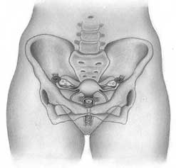
Even considering that most of the female genitals are intrapelvic, the very
typical configuration of the woman's pelvis just by itself lets us know
its content.
It is pertinent to remember here that the very special anatomic features
of the female pelvic bones are partly responsible for the external morphology
of the woman's body from the waist down. Obviously, in addition to this
there is also the typically female distribution of the subcutaneous fatty
tissue on the hips, buttocks and upper parts of the thighs.
Another greatly significant anatomical feature is that through the genitals,
the woman's belly becomes, in a way, "opened" to the exterior.
(Remember that the Fallopian tubes' terminal orifices open directly into
the peritoneal cavity. There is nothing similar in the male genitals.)
The vagina can be seen as the canal or "tunnel" that leads
to the uterus and the interior of the female pelvis. This means that from
the sexual, symbolical and archetypal points of view, it is the "way
in" ( and the "way out" ) of the woman's body. The vagina
possesses a thin muscular layer and is coated by a mucosa lined by a stratified
squamous epithelium highly sensitive to the action of the ovarian hormones.
In repose conditions the vaginal walls remain collapsed, but a typical reaction
of vaginal lengthening and widening takes place during sexual excitement,
culminating with the "ballooning" of the upper third of this organ.
The contractile capacity of the vaginal musculature is very small, and the
strong contractions that take place around the lower third of the vagina
during orgasm does not originate in its muscles but in the muscular
groups of the perineum and pelvic floor that surround the vaginal entrance.
The vulva, in turn, is the "doorway" of the vagina. At the
genital level, the main areas for women's sexual stimulation are found at
the vaginal entrance, the vulvar cleft, the labia minora and majora and,
over and above all, at the clitoris (and, in many women, also at the Gräfenberg
Spot.) (Regarding the G-Spot, which actually is part neither of the vagina
nor the vulva, see my article here at the MUM.)
Behind and around the vulvar structures there are the already mentioned
muscles of the perineum and the pelvic floor. They surround the vaginal
entrance and are capable of giving rise to strong contractions. During orgasm,
they contract rhythmically. Among those that constitute the pelvic floor,
the most important one is the pubococcygeus. In cases of vaginismus, the
psychologically-triggered strong spastic contraction of these muscles is
able to impede sexual intercourse.
Regarding the above mentioned view of the vulva as the female genitals'
"doorway," we must observe that, certainly due to archetypal reasons,
there are amazing morphologic similarities between several doorways of temples,
churches and palaces built by different civilizations of the past and the
vulvar structures.
Just like all the other woman's sexual organs, the trophicity not only
of the vagina but also of the vulvar mucosa and labia is maintained by the
estrogens. |
 |
While the uterus occupies a basically central position inside the female
pelvis, the Fallopian tubes and ovaries are placed bilaterally (see Note 1 below). Though entirely explainable for
reasons of physiological order, this highly "centralized" uterine
position inside the pelvis also seems to be endowed with a particular symbolism
(as to that, see my article "Symmetric Patterns
in the Female Genitals"). In spite of the great mobility of
the uterine corpus (the main part of the womb), the uterus as a whole remains
"anchored" in its basic position by means of the uterine cervix
and an intricate group of ligaments named retinaculum uteri. These
ligaments originate in the uterine cervix and attach to specific points
of the pelvic walls. Of these, the most important ones are the cardinal
and the sacro-uterine ligaments. There are also the pubo-vesico-uterine
ones, but these are not so important as the former.
The cardinal ligaments are bands of fibrous tissue that extend from the
lateral parts of the uterine cervix towards the lateral pelvic walls.
The sacro-uterine ligaments are fibro-muscular cords that, emerging from
the posterior part of the uterine cervix, attach to the walls of the sacrum.
The "anchorage" of the uterus in the pelvis is completed by the
round and the broad ligaments, that arise from the uterine corpus and are
less tense than the others, which provides this part of the organ with its
usual great mobility. The broad ligaments are formed by folds of the
pelvic peritoneum that encase the corporal part of the uterus; they emerge
from the lateral sides of this organ and attach to the pelvic walls. The
broad ligaments give the uterine corpus a curious "winged-like"
appearance. The round ligaments are fibro-muscular cords that arise from
both uterine upper-lateral angles and run to the inguinal canals, terminating
inside the vulvar labia majora. |
|
|
The ovaries are linked to the uterus by means of the utero-ovarian ligaments
and to the pelvic lateral walls through the suspensory-ligaments of the
ovaries or infundibulo-pelvic ligaments; they are also attached to the broad
ligaments through the meso-ovaries. The Fallopian tubes, in turn, are "anchored"
to the broad ligaments through peritoneal folds named mesosalpinges.
By means of this intricate and complex ligamentary system, a direct
anatomic connection between the woman's inner genitals and her pelvic frame
is established. Considering that the morphology of the female pelvic bones
all by itself exhibits typical sexual features, the concept of "pelvis"
in women must be regarded as a entity.
It comprehends everything that, in this part of the body, characterizes
the female sex: vulva, vagina, uterus, Fallopian tubes, ovaries and respective
sustaining structures, pelvic bones and, finally, muscles of the pelvic
floor and perineum. With the addition of its very typical cutaneous, subcutaneous,
fatty and muscular coating on the hips, belly and buttocks, the external
morphology of the female pelvis only by itself is an important element of
sexual attraction, acquiring great importance in the woman's corporal image.
More articles by Dr. Soucasaux in the
column at left.
Note 1:
For more specific details on the uterus, Fallopian tubes and ovaries, see
several other articles of my authorship published here at the MUM ( see
column at left).
Note 2: The text above consist of
excerpts from the introduction of the second chapter of my book "Os
Órgãos Sexuais Femininos: Forma, Função, Símbolo
e Arquétipo" ("The Female Sexual Organs: Shape, Function,
Symbol and Archetype"), published by Imago Editora, Rio de Janeiro,
1993. For more details on the book, see page http://www.nelsonginecologia.med.br/orgaos.htm
, at my website www.nelsonginecologia.med.br
.
Copyright Nelson Soucasaux 1993, 2006 (text and illustrations)
____________________________________________________
Nelson Soucasaux is a gynecologist dedicated to clinical, preventive
and psychosomatic gynecology. Graduated in 1974 by Faculdade de Medicina
da Universidade Federal do Rio de Janeiro, Brazil, he is the author of several
articles published in medical journals and of the books "Novas Perspectivas
em Ginecologia" ("New Perspectives in Gynecology")
and "Os Órgãos Sexuais Femininos: Forma, Função,
Símbolo e Arquétipo" ("The Female Sexual Organs:
Shape, Function, Symbol and Archetype"), published by Imago Editora,
Rio de Janeiro, 1990, 1993. He has been working in his private clinic since
1975.
Web site (Portuguese-English): www.nelsonginecologia.med.br
Email: nelsons@nelsonginecologia.med.br |
|